Summary: This page describes the webmaster’s definition of magnetoencephalography (MEG), the ambiguity of the concept, and the various definitions provided by different experts.
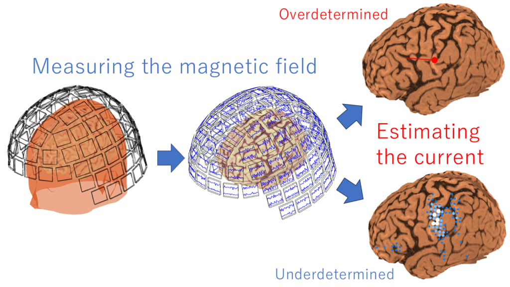
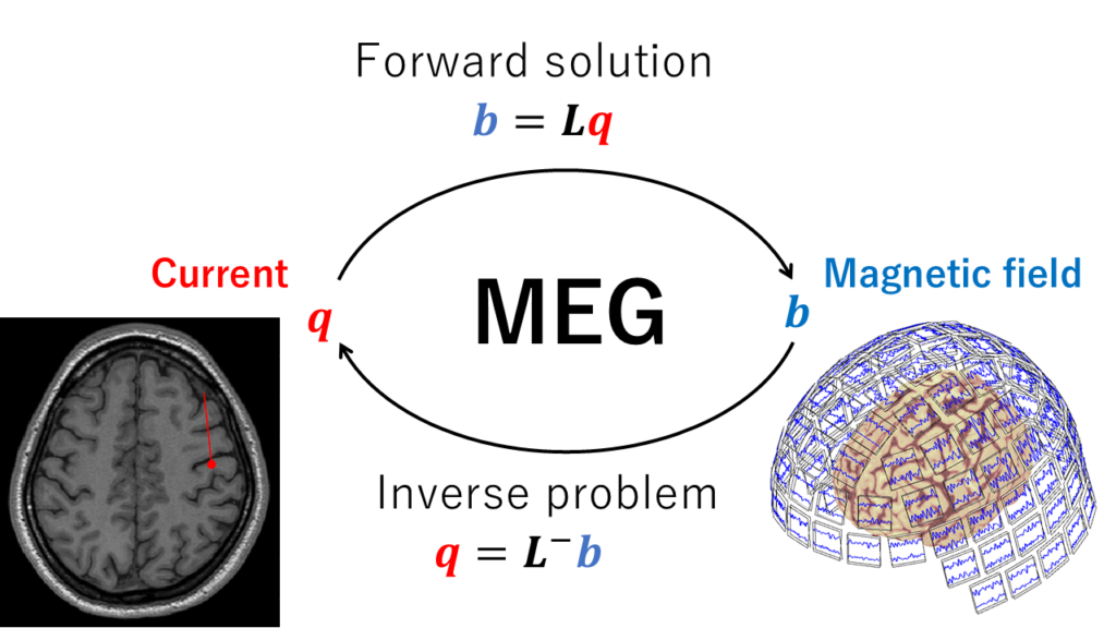
In clinical and neuroscience settings, it is essential to know where and how electrical current is flowing in the brain of a patient or subject in front of us. It would be ideal to acquire this information non-invasively and accurately. Since electrical currents generate magnetic fields, measuring the brain magnetic field allow us to understand the brain electrical current.

The webmaster defines magnetoencephalography (MEG) as follows:
“A methodology to discuss the current flowing in the brain
by measuring the magnetic field the brain emits”

The following section further explores the ambiguity of the concept of magnetoencephalography, as well as the definitions provided by different experts.
Conceptual ambiguity
The term “magnetoencephalography (MEG)” is sometimes used ambiguously.
In clinical practice, the results of MEG analysis are conventionally referred to as MEG, and the general image of that is a representation of a brain with several superimposed current dipoles.
Regarding the concept of magnetoencephalography, it changes grammatically depending on the suffixes -gram, -graph, -graphy:
Magnetoencephalogram: Data or waveform of magnetic output from MEG system
Magnetoencephalograph: MEG system
Magnetoencephalography: Methodology or examination to measure brain magnetic fields
However, in most cases, only the term “magnetoencephalography” is used. In fact, the results of a Google + PubMed search (September 2022) with ” ” yielded the following results:
- Magnetoencephalogram (209,000 + 357 results)
- Magnetoencephalograph (7,300 + 30 results)
- Magnetoencephalography (692,000 + 11,025 results)
It seems that the term “magnetoencephalography” is predominantly used. Although it might be better to use “magnetoencephalogram” when emphasizing it is the waveform and “magnetoencephalograph” when emphasizing it is the measurement device, such distinctions are not widely observed. In particular, except for some companies, “magnetoencephalograph” is rarely used.
To summarize, “magnetoencephalography” is often used to mean:
- The analysis results representing current dipoles superimposed in the brain
- The waveform of magnetic output from the MEG system
- The methodology for measuring brain magnetic fields
Therefore, it is important to be careful not to confuse these meanings.
Definitions provided by various experts:
Experts have varying and inconsistent definitions, although they may seem to indicate similar things. Especially in recent guidelines, the term “magnetoencephalography” is often used without explicit definition.
In the past, the term “SQUID” was included in some definitions. SQUID has been the mainstream and dominant sensor for over 50 years, but it is just one measurement method. Currently, next generation sensors such as optical pumped magnetometers (OPM) and tunnel magneto-resistive (TMR) are emerging.
(Note: SQUID stands for Superconducting Quantum Interference Device. It is a superconducting technology that incorporates Josephson junctions into the circuit, enabling the measurement of extremely fine magnetic fields, down to 10 fT (femtotesla). By combining it with a shield room that blocks external magnetic fields, it became possible to measure the weak brain magnetic fields of humans.)
Here are some representative definitions provided by various experts:
“The magnetic signal generated by the brain is described as brain magnetic field = magnetoencephalogram (MEG). It is also used to mean the methodology of measuring brain magnetic fields (Hiroshi Hara and Shinya Kuriki, eds., Brain Magnetics Science – SQUID Measurement and Medical Applications, Ohmsha, p. 126).”
“An image obtained by recording tiny magnetic signals generated by nerve activity currents using a measuring device (Japan Consortium of Clinical MEG).”
“Magnetoencephalography (MEG) is a neuroimaging tool which obverts functional brain maps or pathognomonic signs with millisecond order. (Shozo Tobimatsu, Ryusuke Kakigi. Clinical Applications of Magnetoencephalograpy. Springer 2016. P.v,3.)”
“A completely noninvasive procedure that uses an array of highly sensitive sensors to detect and record the magnetic fields associated with electrical activity in the brain” (The HP of American Clinical Magnetoencephalography Society).