Abstract: There is a minimum unit for discussable current based on the measurement of magnetic fields by magnetoencephalography (MEG).
In the previous page, the concept of current dipoles was introduced, which represents the current as a vector quantity called the current moment. The unit was stated to be Am. It was also mentioned that current dipoles are point-like and have a position.
This allows for the convenient discussion of brain currents as point currents (without considering the spatial extent of the current) [1].
In the measurement of currents using magnetoencephalography, it is traditionally believed that a minimum unit of 10 nAm is required [2]. This value is not determined experimentally but is based on plotted results of calculations.
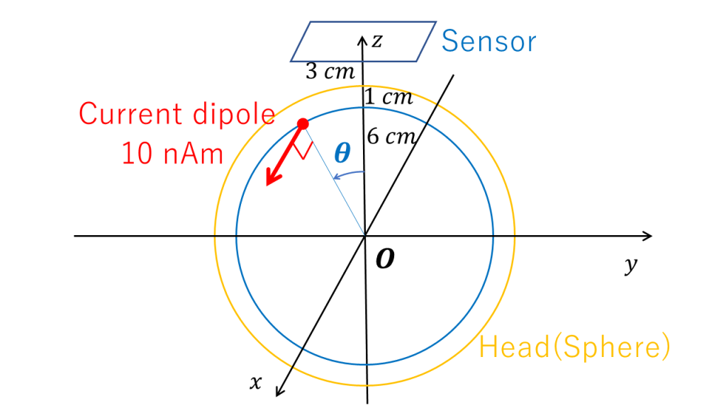
The following figure presents an example of typical measurement conditions, where the administrator’s brain is used for a virtual experiment. The forward problem is solved using the Sarvas formula [1], considering a virtual sphere with a radius of 7 cm representing the brain surface. The sensors are assumed to be fixed horizontally at the center of the virtual sphere, with a distance of 3 cm from the brain surface to the sensors. The baseline distances for planar gradiometers and axial gradiometers are assumed to be 1.68 cm and 3 cm, respectively.
It is assumed that a current of 10 nAm flows in the vertical and horizontal (x-axis) directions on the brain surface at a depth of 1 cm from the brain surface, specifically for planar gradiometers. To observe the sensor’s response, the current is moved. The current dipoles are assumed to move in a half-circle motion, starting from the point (0, 6, 0) relative to the center of the virtual sphere, passing through (0, 0, 6), and ending at (0, -6, 0) in a plane (yz plane) perpendicular to the current.

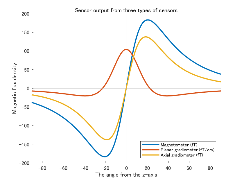
This results in the graph shown below.

In clinical practice, if the waveform can be visualized at around 200 fT or 100 fT/cm, it can be considered as the lower limit of detectability. Therefore, under typical measurement conditions, 10 nAm can be regarded as a detectable unit.
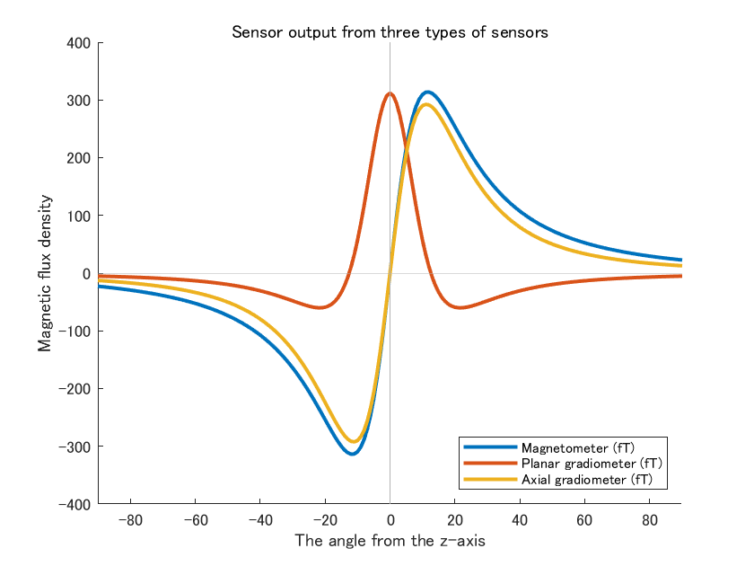
If a graph similar to Fig 27 in Hämäläinen’s review [2] is desired, the following parameters can be used: virtual sphere with a radius of 10 cm, distance from the brain surface to the sensors of 0.25 cm (not mentioned in the text), baseline distances for planar gradiometers and axial gradiometers of 1.36 cm (1.5 cm in the text) and 6 cm, respectively, and a current depth of 3 cm. To obtain the output of planar gradiometers in fT without dividing by the baseline distance, the unit can be converted directly to fT (see the figure below).

However, the distance between the brain surface and the sensors is too close to reality, and as a result, the specific distance between the sensor and the brain surface is not described in the article. Additionally, a brain with a radius of 10 cm corresponds to a volume of 4,200 mL, which is three times the volume of the administrator’s brain (radius of 7 cm). The reason for adopting these numbers is unclear, but it is likely that such virtual situations were assumed to create clear diagrams.
As a side note, from these graphs, it can be observed that in magnetometers and axial gradiometers, the sensor output becomes zero when there is no current intended for measurement directly below the sensor. This also means that in order to locate the position of the current intended for measurement, which forms the positive and negative valleys of the
sensor output, it is necessary to draw equipotential field lines and search for the zero line.
The webmaster believes that such complex behavior of sensor outputs has hindered the intuitive understanding of magnetoencephalography and deterred beginners.
Summary: The minimum unit required for measuring currents using magnetoencephalography is achievable under typical measurement conditions.
In the next page, I would like to discuss the surface area of the cerebral cortex required to generate a minimum current moment of 10 nAm.
(References)
- J Sarvas. Basic mathematical and electromagnetic concepts of the biomagnetic inverse problem. Phys Med Biol. 1987 Jan;32(1):11-22.
- MS Hämäläinen, R Hari, RJ Ilmoniemi, J Knuutila, OV Lounasmaa: Magnetoencephalography theory, instrumentation, and applications to noninvasive studies of the working human brain. Rev Mod Phys. 1993 Apr;65(2):413-97.